General Methods for Cell Apoptosis Assay
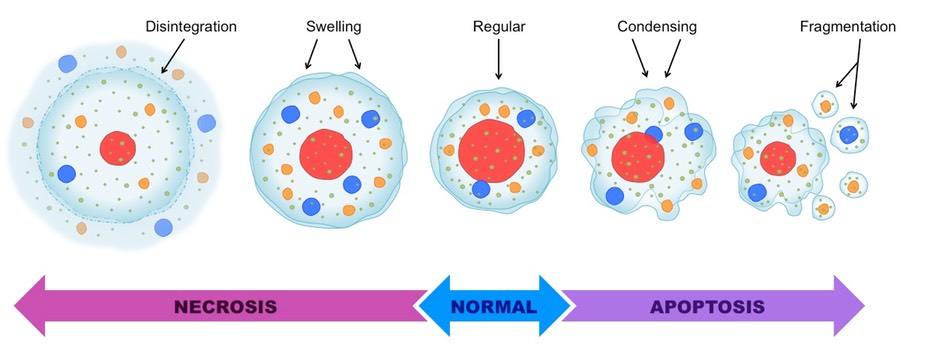
What is cell apoptosis? Cell apoptosis refers to the programmed cell death controlled by genes to maintain stable internal conditions. Differing from cell necrosis, apoptosis is an active process which involves the activation, expression, and regulation of a series of genes. instead of the passive one. It is not a form of traumatic cell death that results from acute cellular injury, apoptosis is a highly regulated and controlled process that confers advantages during an organism's life-cycle. According to the difference in morphology, biochemistry, and molecular biology of dead cells, it will be easy to distinguish cell apoptosis and cell necrosis. Some commonly used cell apoptosis assay methods: 1.Morphological detection of cell apoptosis A.Optical Microscope and inverted microscope to detect the unstained cells and stained cells. B.Fluorescence microscope and Laser scanning confocal microscopy C.Transmission Electron Microscope 2.Annexin V Phosphatidylserine (PS) is normally located inside the cell membrane, but at the early stage of cell apoptosis, PS can be flipped from the inside of the cell membrane to the surface of the cell membrane and exposed to the extracellular environment (Figure 3). Annexin-V is a Ca2 + -dependent phospholipid-binding protein with a molecular weight of 35-36KD, which binds specifically to PS affinity. Annexin-V was labeled with fluorescein (FITC, PE) or biotin. Taking advantage of Annexin-V that has been labeled with fluorescein (FITC, PE) or biotin to detect the occurrence of apoptosis by flow cytometry or fluorescence microscopy. 3.Detection of Potential Energy of Mitochondrial Membrane Mitochondria play a pivotal role in the process of apoptosis, a variety of apoptosis stimulating factors can induce apoptosis of different cells, and mitochondrial transmembrane potential DYmt decline, is considered as the earliest occurrence in the process of cell apoptosis cascade, which occurs when the nucleus apoptosis feature (chromatin condensation, DNA cleavage) occurs, once the mitochondrial DYmt crashes, the apoptosis will be irreversible. 4.DNA fragmentation detection The main biochemical characteristic of cell apoptosis is that its chromatin is concentrated and chromatin DNA breaks at the junction between nucleosomal units to form a large fragment of DNA from 50 to 300 kbp or an integer multiple of 180 to 200 bp Acid fragments, performing as ladder electrophoresis (DNA ladder) in the gel electrophoresis. After the cells were treated, the purified DNA was isolated and purified by conventional methods, stained with agarose gel and ethidium bromide, and typical DNA ladder was observed in the apoptotic cell population. If the amount of cells is small, DNA can also be labeled with 32P-ATP and deoxyribonucleotide terminal transferase (TdT) after separation and purification of DNA, followed by electrophoresis and autoradiography to observe the presence of DNA ladder in apoptotic cells. 5.TUNEL (terminal -deoxynucleotidyl transferase mediated nick end labeling) In the process of apoptosis, the chromosomal DNA double-strand breaks or single-strand breaks produce a large amount of viscous 3'-OH terminus, which can react with deoxyribonucleotides and fluorescence under the action of deoxyribonucleotide terminal transferase (TdT) Derivatives of peroxidase, detecting apoptotic cells by labeling alkaline phosphatase or biotin to 3'-terminus of DNA, that is TUNEL. TUNEL is actually a combination of molecular biology and morphology research methods, the complete single apoptotic nuclei or apoptotic bodies in situ staining, can accurately reflect the typical apoptotic biochemical and morphological characteristics. 6.Expression and cell localization of apoptosis - related protein TFAR19 The expression level and localization of TFAR19 protein in the early stage of apoptosis were observed by using the TFAR19 monoclonal antibody labeled with fluorescein (FITC) as the probe. The expression of TFAR19 in the early stage of apoptosis increased and the rapid nuclear translocation was observed accompanied by changes in the morphology of the nucleus, which lasts longer and remains visible in apoptosis body. All the mentioned above is about the general methods which can be used to detect cell apoptosis. Except for these methods, there are still some methods are available, such as the Western blot analysis, Fluorescence spectrophotometer analysis and Flow cytometry analysis etc. Related service: Cell apoptosis
Your email address will not be published. Required fields are marked *