Biochemical 3D Vascularized Tissue Model - Improving The Accuracy of Drug Toxicity Tests
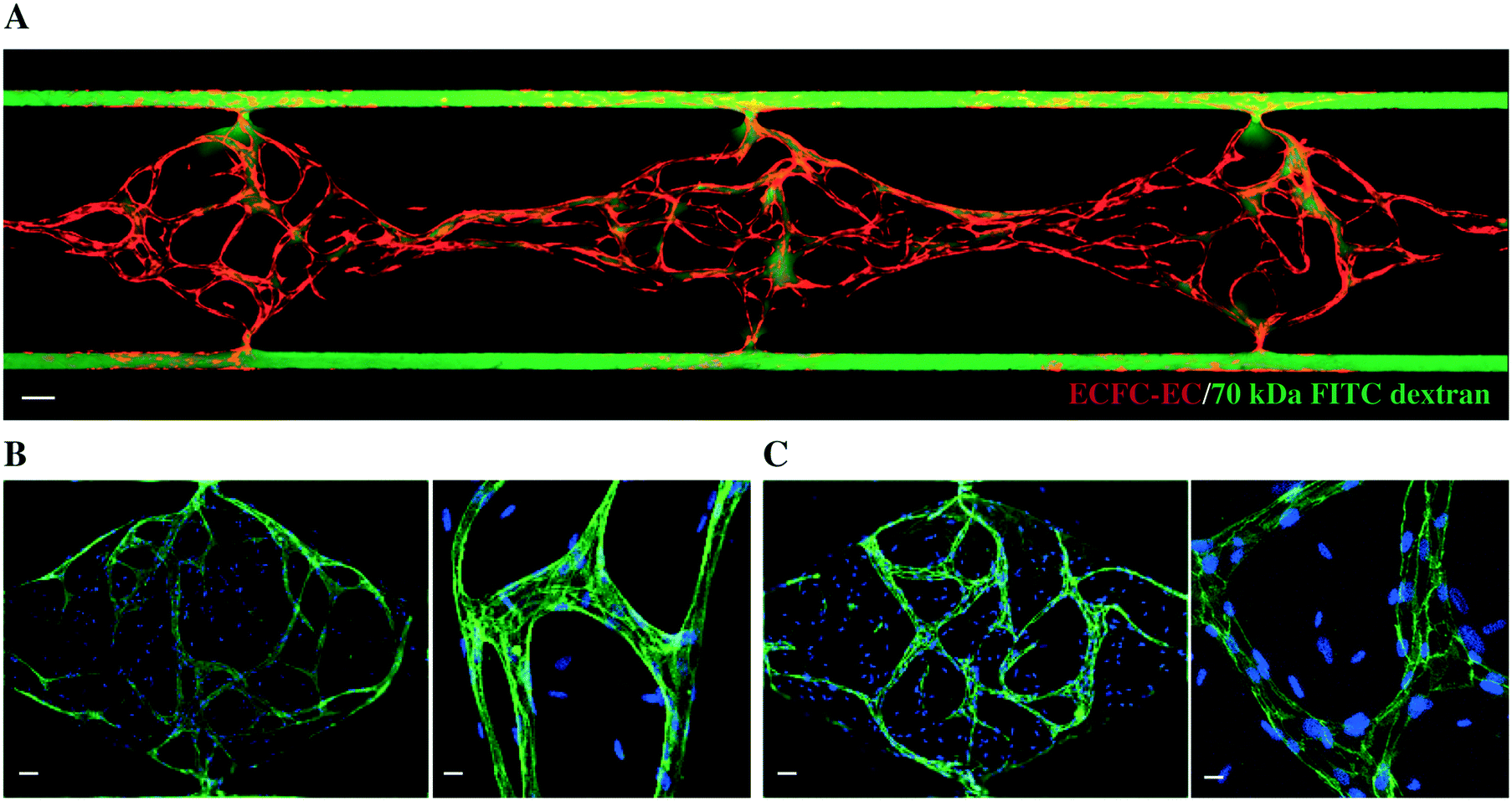
Researchers from Chile, Italy, Saudi Arabia, South Korea and the United States have recently printed out a simple vascularized liver tissue by collaborating with 3D biology. The study has been published in the journal Biomicrofluidics, entitled as "Biomedical 3D vascularized tissue model for drug toxicity analysis". This breakthrough may have important applications in the field of drug toxicity testing and drug screening. The print material is a special "sacrificial" biological ink (a material that can be washed away), and the final 3D blood vesselized liver tissue model can be used for more accurate drug toxicity testing. Researchers even claimed that their 3D bioprocessing methods could also be used to print different types of cells, which brought more possibilities for personalized drug screening. "Most drug test models use a 2D monolayer cell tissue or a nonvascularized 3D tissue," the researchers explained, "however, our body is made up of a variety of vascularized 3D tissues, not just monolayer cells." With the use of sacrificial biological inks, researchers can mimic the structure and function of human tissue and successfully print out tissues with complex microchannel stents. Compared with 2D monolayer cell modules, the use of 3D structures makes drug toxicity testing more accurate. Although this biological print of liver tissue is relatively simple at this stage, but the researchers still made some important discoveries, including the drug diffusion response being delayed by the organization's endothelium. For the further study, researchers expect that they can eventually be able to print out more complex drug testing systems with less time, such as "multiple organs on a chip" device or other organ tissue. Ultimately, they hope that this project will help promote the development of cancer drug therapy and other fields.


Your email address will not be published. Required fields are marked *