
The CCK-8 Protocol Guide: Principle, Step-by-Step, and the 3 Mistakes That Ruin Your Data
Cell Counting Kit-8 (CCK-8) has become the gold-standard colorimetric method for assessing cell proliferation and cytotoxicity. Built on the water-soluble tetrazolium salt WST-8, it offers superior sensitivity, minimal toxicity, and a streamlined workflow compared to legacy assays like MTT. Yet even experienced researchers can compromise their data with subtle protocol deviations.
This guide breaks down the CCK-8 principle, provides a reproducible step-by-step protocol, and highlights the three most common pitfalls that undermine assay reliability—along with actionable solutions to ensure your OD readings at 450 nm truly reflect cellular activity.
What Is the CCK-8 Assay?
CCK-8 (Cell Counting Kit-8) utilizes WST-8, a water-soluble tetrazolium salt that is reduced by mitochondrial dehydrogenases in viable cells to produce an orange formazan dye. The intensity of the color is directly proportional to the number of living cells, making it ideal for both proliferation and cytotoxicity studies.
Unlike MTT, the resulting formazan is water-soluble—eliminating the need for toxic organic solvents like DMSO and enabling continuous monitoring without cell lysis.
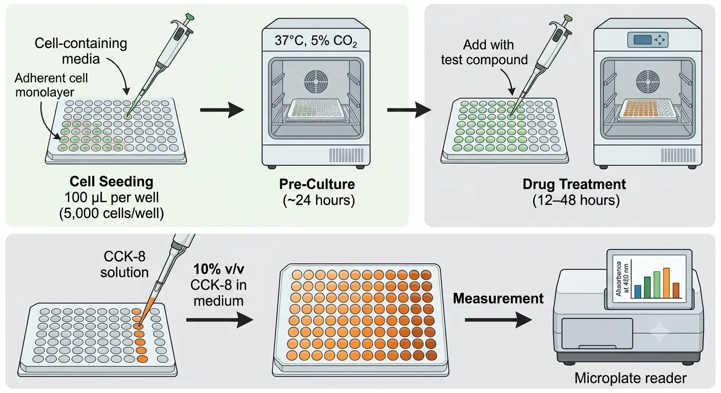
Step-by-Step Protocol
1. Cell Seeding
Harvest cells in logarithmic growth phase. For adherent cells, digest with trypsin, neutralize with serum-containing medium, and centrifuge. Resuspend in complete medium and adjust to 5 × 10⁴ cells/mL. Add 100 µL per well to a 96-well plate (final density: 5,000 cells/well). Include 3–6 replicate wells per condition, plus a cell-free blank and a vehicle-only control.
2. Pre-Culture
Incubate the plate at 37°C, 5% CO2 for ~24 hours to allow cells to adhere and enter steady-state growth.
3. Drug Treatment
Replace medium with fresh medium containing the test compound at desired concentrations. Return to the incubator for 12–48 hours, depending on the mechanism of action.
4. CCK-8 Addition
Add 10 µL of CCK-8 solution per 100 µL of medium (10% v/v). To minimize pipetting error and residue on tips, pre-dilute CCK-8 in culture medium and mix thoroughly before dispensing. Gently swirl the plate to distribute the reagent evenly, avoiding bubble formation.
5. Color Development
Incubate at 37°C for 1–3 hours. Monitor color change periodically; the solution should turn orange. Aim for an OD450 between 1.0 and 3.0 to remain within the linear detection range.
6. Measurement
Read absorbance at 450 nm using a microplate reader. If turbidity is high, use a reference wavelength of 600 nm or higher and subtract background noise.
Critical Pitfalls & How to Avoid Them
Pitfall 1: Incorrect Seeding Density & Edge Effects
The Risk: Seeding too few cells pushes signals below the detection threshold, while over-seeding saturates the reader. Meanwhile, evaporation in the outer perimeter wells of a 96-well plate introduces volume variability and skewed OD values.
The Fix:
- Maintain a minimum of 1,000 adherent cells per 100 µL well to ensure reliable signal generation.
- Fill the outer ring of the plate with PBS only and exclude these wells from analysis. This eliminates edge-effect evaporation and improves inter-well consistency.
Pitfall 2: Ignoring Drug Interference & Blank Controls
The Risk: Certain compounds absorb light at 450 nm or chemically interact with WST-8, producing false-positive or false-negative results.
The Fix:
- Always run a cell-free blank containing medium, drug, and CCK-8 (no cells). Subtract this value to correct for drug-specific absorbance.
- Include a vehicle control (cells + medium + CCK-8, no drug) to establish baseline proliferation.
- Use the validated formula:
Pitfall 3: Suboptimal Incubation Time & Reagent Handling
The Risk: Under-incubation yields weak signals; over-incubation drives OD outside the linear range. Old or improperly stored CCK-8 reagent develops elevated background, while careless pipetting introduces bubbles that scatter light.
The Fix:
- Optimize incubation time empirically. Check the plate every 30 minutes until the OD450 falls within 1.0–3.0, then lock that duration for all replicate experiments.
- Store CCK-8 at 4°C protected from light for up to 6 months, or at -20°C for up to 1 year. Fresh reagent is pink; darkening indicates degradation.
- Pipette slowly against the well wall to prevent bubbles. If bubbles appear, briefly centrifuge the plate or use a fine needle to puncture them before reading.
CCK-8 vs. MTT: Why Make the Switch?
The CCK-8 assay's single-bottle format, low toxicity, and compatibility with downstream assays make it the preferred choice for high-throughput drug screening and longitudinal proliferation studies.
| MTT | CCK-8 | |
|---|---|---|
| Formazan Solubility | Insoluble (requires DMSO) | Water-soluble |
| Workflow | Multiple steps, cell lysis | Single-step, non-destructive |
| Sensitivity | High | Higher |
| Cytotoxicity | High | Very low |
| Detection Time | Longer | Shortest |
| Stability | Normal | Excellent |
Accelerate Your Research with Professional Cell Assay Services
While an optimized in-house protocol is invaluable, complex drug-screening campaigns and large-scale cytotoxicity studies often demand higher throughput and specialized expertise. Creative Bioarray offers comprehensive cell viability and cytotoxicity assay services, including validated MTT/CCK-8 method testing performed by experienced technicians under GLP-compliant workflows.
Whether you need high-throughput 96/384-well microplate screening, custom 3D spheroid cytotoxicity models, or simply a reliable Cell Counting Kit-8 reagent for your own lab, Creative Bioarray provides end-to-end support—from cell culture and compound treatment to image collection and quantitative data analysis. Outsourcing your CCK-8 assays can free up internal resources, reduce batch-to-batch variability, and deliver statistically powered datasets faster.
Ready to eliminate assay variability? Explore Creative Bioarray's Cell Viability, Proliferation and Cytotoxicity solutions and let our team handle the technical pitfalls so you can focus on the science.
Your email address will not be published. Required fields are marked *