
Organoid Budding, Branching, and Fusion: What Do They Really Mean?
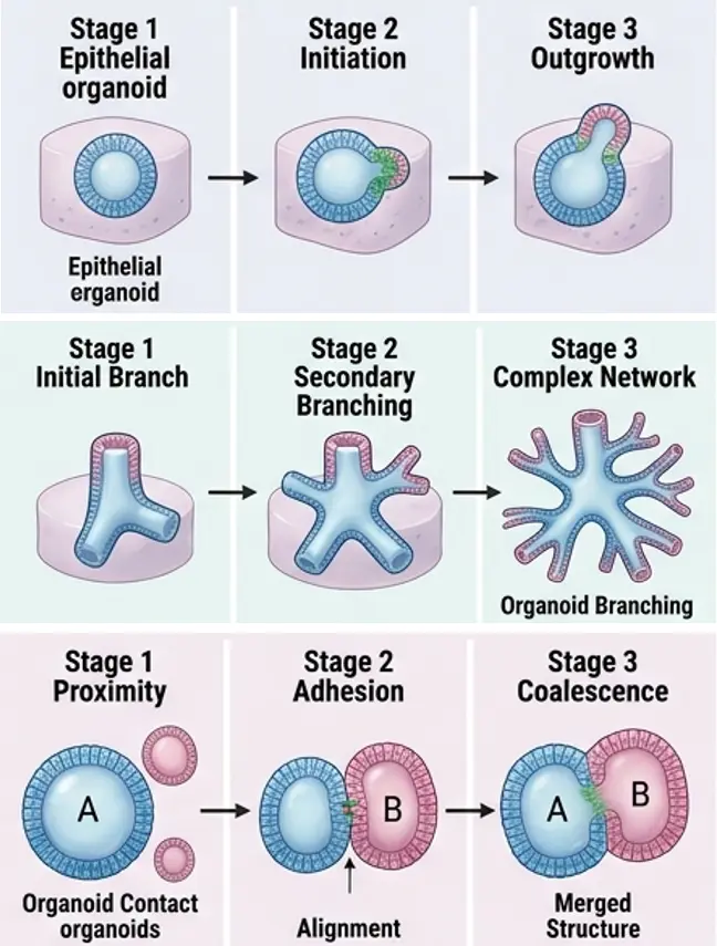
An outward bud, a branching extension or two organoids joining into one structure can make a culture look more elaborate. Yet appearance alone cannot establish differentiation, functional maturity or model quality. Interpretation depends on the organ being modeled, the protocol, the culture stage and the phenotype the experiment is designed to measure.
Budding, branching and fusion are different processes
| Dfinition | Question to ask | |
|---|---|---|
| Budding | A localized protrusion grows outward from an existing structure. It may develop further or remain transient. | Does the protrusion have organized cells, polarity and a reproducible growth trajectory? |
| Branching | An extension establishes a discernible new growth axis, potentially producing further branch points and tips. | Is the branch connected, stable and appropriate for this particular model? |
| Fusion | Previously separate structures meet and become physically continuous. | Did contact simply merge masses, or did the tissues integrate in an organized, testable way? |
These are descriptive terms, not a universal sequence of increasing maturity. A bud can precede a branch in some systems, while fusion involves contact between structures and has a different origin. Defined assembloid experiments deliberately bring distinct populations together to study their interactions; accidental coalescence in a crowded well does not carry the same interpretation.
What do these shapes actually tell you?
Buds: localized growth needs context
In a suitable epithelial model, a bud can reflect patterned proliferation, cell shape changes and rearrangement of polarity. But a protrusion can also accompany an altered matrix, uneven growth factor exposure or disorganized expansion. Follow the same structure over time. Ask whether the bud persists, elongates, maintains a coherent epithelium and expresses the expected markers. A large count of irregular protrusions is not itself evidence of improved differentiation.
Branches: model-specific architecture matters
Branching morphogenesis is a feature of developing organs such as the lung and mammary gland, and organoid systems can be designed to model aspects of it. In a primary mammary organoid study, changes to the extracellular matrix and timed growth factor delivery supported primary, secondary and tertiary branches—an example of why culture conditions must be recorded alongside morphology. The developing kidney also branches through the ureteric bud, but this does not mean every kidney organoid protocol should form a branched collecting duct tree. Judge the result against the specific tissue compartment and protocol, not the organ name alone.
Useful measures include the fraction of organoids with branches, branch length, tip count, branch order and lumen continuity. Define how each measure is scored before comparing groups, and report the distribution across independent cultures rather than displaying only a representative image.
Fusion: contact is not the same as integration
When two organoids are seeded near each other, growth or movement may bring them into contact. The disappearance of a visible border establishes neither a new tissue identity nor a new function. For an intentional fusion or assembloid experiment, examine spatial organization of each starting population, continuity across the interface, cell interactions and a relevant functional outcome. For unexpected widespread fusion, first inspect seeding density, initial fragment size and how the matrix separates structures.
A practical framework for validating morphology
- Define the expected phenotype. State the organ compartment, developmental stage, protocol and purpose of the assay. Specify whether budding, branching or integration is an intended endpoint.
- Capture the trajectory. Image the same structures at suitable intervals, using consistent magnification and segmentation rules. Distinguish a persistent branch from a temporary bulge or structures that happened to touch.
- Quantify architecture and viability. Record size, growth, lumen organization, protrusions, branch tips and the fraction of fused structures, as appropriate. Assess whether larger structures have poorly viable cores; size can alter nutrient and oxygen gradients.
- Verify identity and organization. Use markers suited to the model for lineage, proliferation, polarity and cell death, with spatial information where possible. Ki-67 positivity alone shows proliferation, not correct developmental patterning.
- Test relevant function. Choose an assay tied to the question—such as barrier integrity, secretion, transport or treatment response. Interpret it together with morphology and identity, rather than assuming shape predicts function.
Quantitative morphology improves consistency, but there is no single circularity or branching threshold that applies to all organoids. Published quality control work emphasizes multidimensional, model-specific assessment and reproducibility.
When the culture suddenly changes shape
More budding or branching than expected? Compare time courses with a reference batch. Then check starting fragment size and passage history, matrix lot and gelation, embedding geometry, medium preparation and growth factor exposure. Change one plausible variable at a time where feasible. A new branch pattern may reflect altered signals or mechanics; it should not be labeled “better” before cell identity and function are checked.
More fusion than expected? Review structures per well, their spacing and size, handling during embedding, and matrix integrity. Track which structures were separate at baseline. If the experiment aims to create an assembloid, label the input populations and test the interface. If it aims to compare individual organoids, record and manage fusion as an assay variable rather than silently pooling those structures with isolated ones.
Why this distinction matters in drug screening
A compound may change proliferation, adhesion, polarity, matrix remodeling or viability, any of which can alter an organoid’s outline. More buds after treatment therefore do not automatically mean enhanced growth, and a larger fused mass is not automatically a healthier organoid. Predefine morphology readouts, include vehicle and culture controls, and pair images with an orthogonal measure of the biological effect under study. Keep the unit of analysis clear: a merged structure is not equivalent to two independent replicates.
Make morphology an interpretable readout
Budding describes an outward protrusion, branching describes the establishment of new growth axes, and fusion describes continuity between previously separate structures. Their value lies in a reproducible link between shape, trajectory, cell identity and function. Define that link for each model before treating a striking image as a biological conclusion.
Build a clearer organoid workflow
Reading morphology correctly depends on controlling everything upstream of the image: the matrix, the medium, the seeding strategy, and the assay readout. Creative Bioarray supports 3D culture and organoid research with a portfolio designed for consistency and scale, including:
- Organoid culture media and reagents formulated to reduce lot-to-lot variability in growth factor composition.
- Extracellular matrix and 3D culture consumables for reproducible gelation and spatial separation between structures.
- Patient-derived organoid models and custom organoid development for disease modeling and drug response studies.
- Functional assay and screening services covering viability, imaging-based phenotyping, and mechanism-oriented readouts.
- Technical consultation on culture parameters, quality-control design, and quantitative morphology workflows.
Whether you are troubleshooting unexpected budding, validating a branching model, or standardizing fusion metrics for screening campaigns, our team can help you turn morphological observations into defined, quantifiable, and reproducible data.
Explore organoid solutions and 3D culture products at https://www.creative-bioarray.com/applications/3d-spheroid-organoid-culture.htm
Your email address will not be published. Required fields are marked *