Co-Culture: A Beginner's Guide to Getting It Right
Single-cell culture struggles to simulate the complex cellular microenvironment in vivo. Cell co-culture technology provides a crucial tool for understanding cell-cell interactions and reconstructing physiological and pathological processes. This technology places two or more different cell types in the same culture system, allowing them to interact in a shared environment, making it a core tool for studying signal transduction, cell growth and differentiation, and the tumor microenvironment.
1. Direct Co-culture: Cell-to-Cell Interaction Studies
Direct co-culture involves directly seeding two or more cell types into the same culture vessel, allowing the cells to interact through direct contact. This method is best suited for studying biological processes dependent on direct cell-cell contact, such as the direct killing effect of immune cells on tumor cells and signal transduction mediated by cell adhesion molecules.
Its core advantage lies in its ability to realistically simulate biological responses triggered by physical contact between cells. The experimental design is simple and direct, allowing for direct observation of the interaction patterns between cells. However, its shortcomings are also significant: after culture, different cell types tend to mix, making it difficult to separate them for subsequent molecular biological analysis, and requiring advanced cell sorting techniques.
2. Transwell Co-culture: Indirect Effects Mediated by Secretory Factors
Transwell co-culture uses a porous membrane insert to divide the culture system into two layers, with different cell types seeded in each layer. The porous membrane allows small secretory factors and signaling molecules to pass freely but prevents direct cell contact, making it specifically designed for studying indirect interactions mediated by secretory products between cells.
This method is widely used in studies of paracrine regulation of tumor cells and stromal cells, cell signal transduction mechanisms, etc. Its greatest advantage is avoiding cell contamination, facilitating subsequent isolation of different cells for targeted analysis, and resulting in higher specificity and accuracy of experimental results. However, its limitations lie in its inability to simulate the biological effects induced by direct cell contact, and it has strict requirements on the pore size of the porous membrane.
3. 3D Co-culture: Functional Simulation in a 3D Microenvironment
3D co-culture involves seeding cells in a three-dimensional scaffold or matrix, allowing the cells to grow and interact within a three-dimensional spatial structure. It is currently the co-culture method that most closely resembles the in vivo tissue microenvironment. By constructing a three-dimensional structure, cells can exhibit morphology, arrangement, and functional states more closely similar to those in vivo, making it suitable for complex research scenarios such as tissue regeneration, tumor microenvironment modeling, and organ-like structure culture.
Its core advantage is its ability to maximally replicate the three-dimensional growth environment of cells in vivo, more realistically reflecting the spatial interactions between cells and the functional performance at the tissue level. However, it has a high technical threshold, with strict requirements on the material selection of the three-dimensional scaffold and the nutrient supply of the culture system, and the experimental cost is significantly higher than traditional two-dimensional co-culture.
4. Conditioned Culture Medium Co-culture: Targeted Study of Secreted Factors
Conditioned culture medium co-culture uses the supernatant (after processing to remove cells and impurities) from one type of cell culture as the culture medium for culturing another type of cell, specifically to study the effects of soluble factors secreted by the former on the latter. This method is the simplest to operate, requires no special culture equipment, and is suitable for preliminary screening of the biological functions of secreted factors.
However, its limitations are also significant. It cannot simulate direct cell-cell contact or interactions in complex microenvironments, and secreted factors may degrade during culture, making it difficult to maintain a stable concentration. Therefore, it is only suitable for exploratory experiments or preliminary validation.
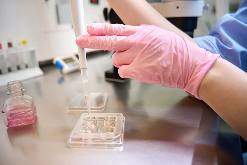
Transwell Co-culture Protocol
Transwell co-culture is the most commonly used co-culture method due to its wide applicability and reliable results. The following is a detailed experimental procedure using the co-culture of tumor cells and fibroblasts (NIH 3T3 cell line) as an example:
Preparation of Experimental Materials
For cell selection, it is necessary to clearly identify the two cell lines to be co-cultured, ensuring that the cells are stable and free from contamination; for Transwell inserts, a 0.4μm pore size is preferred to prevent cell penetration and allow only secreted factors to pass through (8μm pore size is commonly used in migration and invasion assays, and needs to be adjusted according to the experimental purpose); the culture medium should be a type that both cell lines can adapt to, and should be changed to a uniform culture medium in advance to avoid affecting cell growth due to nutritional differences.
Fibroblast Seeding (Lower Chamber Seeding)
First, select fibroblasts with a confluence of 70%-80%, digest them with trypsin, centrifuge them, resuspend them in culture medium, and count the cells. Then, seed the fibroblasts into the lower chamber of a Transwell plate (commonly 6-well or 24-well plates). The recommended seeding density for a 6-well plate is 4 × 10⁵ cells/well, ensuring even coverage of the bottom of the well. After seeding, incubate the plate at 37°C in a 5% CO2 incubator for 4 hours until the cells are fully adhered before seeding the upper chamber to prevent cell detachment during subsequent operations.
Tumor Cell Seeding (Upper Layer Seeding)
Select tumor cells with a confluence of 70%-80%, digest, centrifuge, resuspend, and count them. Using a pipette, vertically suspend the tumor cell suspension and drop it evenly onto the upper layer insert of the Transwell. The plating density needs to be adjusted according to the experimental purpose, with a general range of 5 × 10⁴ -2 × 10⁵ cells/insertion. During seeding, avoid generating air bubbles to prevent affecting cell adhesion and nutrient exchange.
Establishment of the Co-culture System
Carefully place the Transwell insert containing tumor cells into the lower layer culture plate containing fibroblasts, ensuring full contact between the insert and the lower culture medium without leakage. Place the entire co-culture system in an incubator and maintain culture conditions of 37°C and 5% CO2. The culture time is set according to the experimental purpose; for the first experiment, it is recommended to set time gradients such as 24 hours and 48 hours to observe differences in cell interactions at different time points.
Experimental Endpoint Analysis
After co-culture, various assays can be performed: at the cellular level, proliferation, migration, and invasion assays can be conducted to observe changes in cell morphology; at the molecular level, cellular RNA or proteins can be extracted to detect changes in related gene expression or protein levels; culture supernatant can also be collected to analyze changes in the types and concentrations of secreted factors, comprehensively elucidating the mechanisms of cell-cell interactions.
Outsource Cell-Based Assays With Confidence
Our portfolio of cell-based assays use luminescence or fluorescence detection to monitor various cellular events, helping you build a complete picture of how and why cells die. All of our assays use a standard plate reader for detection, and some are also capable of live-cell kinetic measurement or have been optimized for use in 3D cell culture.
Your email address will not be published. Required fields are marked *