What are Epithelial Cells & How to Culture Epithelial Cells
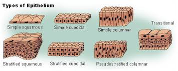
Definition Epithelial cells are cells located on the surface of the skin or cavity. Epithelial cells vary depending on the organ: Urinary routine epithelial cells are decaying, shedding skin cells. Epithelial tissue is divided into coated epithelium, glandular epithelium and sensory epithelium, and epithelium usually referred to the coating epithelium. 1. Epidermal cell culture A. Sample preparation: cutting skin graft into small pieces with 0.5 to 1 square centimeters. B. EDTA treatment: Placing these small pices into 0.02% EDTA at room temperature for 5 minutes. C. Cold digestion: putting into 0.25% trypsin at 4 ℃ overnight. D. Separation: To separate the skin from the dermis with vascular clamps or tweezers. E. Warm digestion: cutting skin into smaller pieces with scissors, and then putting into the new 0.25% trypsin at 37 ℃ and then digested for 30 to 60 minutes. F. Blowing with straw gentally in order to make cell suspension. G.Culture medium: filtering with 80 mesh stainless steel mesh, low-speed centrifugation, suction supernatant, and then placing it directly into the Eagle solution and 20% calf serum to make cell suspension, and inoculating into the dish, culturing in CO2 incubator. 2.Gastric epithelial cell culture A. Sample preparation: Gastric ulcer or gastric cancer surgical removal of gastric specimens B. Cleaning: Rinsing with gentamicin (400 μg / ml) and amphotericin (2 μg / ml Hanks), and peeling the mucosa with a blunt body and cutting into 1 mm size. C. Digestion: Digesting in type I collagenase and hyaluronidase at 37 ° C for 80 minutes. D. Centrifuge: Collect cell suspension, 800 rpm after centrifugation, Hanks solution rinses twice. E. Inoculation: After the last centrifugation, adding to the complete medium containing 1% ~ 2% fetal bovine serum, and then inoculating into different culture plates, the inoculation amount is according to different experimental purposes. 3.Endothelial cell culture A. Sample preparation: The fresh umbilical cord with 10 to 15 cm; other large blood vessels such as embryos and larvae can also be used for culture. B. Injecting PBS with three-way syringe suction into the vein of the umbilical cord. And then injecting the final concentration of 0.1% of the collagenase into umbilical vein slowly. After tying both ends, digesting for 3 to 10 minutes. C. Aspirating the digestive solution containing endothelial cells into the centrifuge tube. D. Absorbing the supernatant, adding 1640 medium to make cell suspension, inoculating into the bottle culture, cells can grow into a single layer within 2 to 3 days. These are some basic epithelial cells culture experiemental instruction and some basic information about it. Related products: Epithelial cells Human Umbilical Cord Cells


Your email address will not be published. Required fields are marked *