The Cell Function Evaluation in Biofunctional Screening and evaluating
At present, Evaluations of cell-related function are divided into independent experimental techniques through existing drug development literature and information. In this post, we aim to systematize the experimental techniques related to these cells and systematically classify the cellular functional assessment into four categories according to the multistage process of cellular response after the signal is received. In addition, we will summarize the main experimental techniques for the evaluation of the four functional categories. The appropriate functional evaluation methods for various drug development phases such as basic research, drug target validation, drug screening, functional evaluation and quality control are discussed. Furthermore, taking the functional evaluation method of G-protein coupled receptor and ADCC as an example, the current mainstream technology of cell drug screening evaluation is introduced to discuss the basic principles of methodology development.
Overview of the cellular physiological processes
Cell is the basic unit of life activity and the functional evaluation method that taking cells as research object is different from the biochemical reaction(protein-protein interaction and enzyme-catalyzed reaction) under the ideal conditions in vitro. The physiological response to the process is multi-stage, may involving complex process such as multiple receptor protein/signaling pathway, Cooperation/resistance.
Single cell physiological response process can be divided into four aspects:
1.Intracellular proteins or membrane receptors bind to outside molecular at the cell membrane, receiving signals from the outside.
2.The protein or receptor that receives the signal changes in its conformation, polymerizes or changes its ability to bind to other proteins in the cell, and signals are transduced through second messengers or kinases, and amplified step by step.
3.The amplified signal continues to affect the downstream transduction pathway, affecting the activity of the transcription factor, which enters the nucleus, mediates transcription of the Nucleic acids, and transcribes the RNA into the cell matrix for regulatory protein expression.
4.The Abundance of the protein expression leads to a final response of the cell to the signaling molecule, causing the cell to excrete material, cell proliferation or death.
 The physiological reaction process of cells after receiving the external signal is often not a single, may involve multiple signal pathways, the complex process can not make a deterministic distinction just based on the literature research. This requires researchers to comprehensively evaluate the changes in the activity and concentration of key markers in cell-receiving signals, signaling, and response to the entire physiological response and confirm the specific signaling pathways through different experimental methods and experimental results. Besides, researchers also need to figure out whether the single cell evaluation screening model established in the process of complex physiological reaction is suitable and can serve the subsequent drug development, especially the new drug research and development, and achieve the expected purpose of use.
 Classification of cell functional assessment methods
According to the single physiological reaction process, the cell functional assays can be divided into the following four categories:
1.Determining the initial alteration of the target protein by the candidate drug, the most common is the binding of a candidate drug to a target receptor located on the cell membrane or the binding of a target receptor to a natural ligand, and commonly used assays that include cell-based ELISA and flow cytometry can be used to determine candidate drug-receptor conjugates. Differ from the target receptor recombination purified affinity evaluation, the target receptor on the cell membrane is closer to the true physiological conditions, the other receptors and the effector protein on the cell may be involved in the activation of the target protein changes, and including more reference value. Moreover, many membrane proteins are multi-permeable membrane structures. It is difficult to purify the receptor proteins that have a complete tertiary structure conformation (eg, G-protein coupled receptors are membrane-permeable).
2.Determination on the concentration of key biomarkers in signaling pathways during signal transduction, such as cAMP, DAG, IP3 (TR-FRET, ELISA) and Ca+2 (FLIPR) during GPCR activation; key effect proteins phosphorylation and other modified concentrations (TR-FRET, WB); concentration determination of key enzyme; kinetic transport process (internalization) etc.
3.Determination of the level of transcription and translation that cause cellular responses. For example, RT-qPCR was used to measure the level of transcription RNA. The transcription factor activity was measured by inserting a foreign reporter gene behind the promoter.
4. Cell proliferation or death was measured by MTT, CCK-8, Alarmable, ATP assay, LDH assay, proteomic or genomic changes of the cells (flow cytometry cycles of apoptosis), cell migration and migration capacity changes (scratch, invasion, transwell experiments), determination of cell exudates such as TNF and IL quantification of PBMC cells (ELISA, TR-FRET).
All the mentioned above is the brief summary of cell function evaluation methods, from which we can lean that there are various methods can be applied to evaluate cells function, but how to choose the appropriate test method to satisfy the research purpose still is a key point. Researchers already shared several suggestions on choosing an appropriate method:
1.In the initial stage of basic research and target identification of drug development. Scientists should combine with the literature and process basic functional and functional experiments at various stages of the cell's single physiological response process, that is signal reception (target protein binding), signal amplification (second messenger or kinase assay), transcriptional translation (RT-qPCR or reporter gene system), final response (cell viability assay, secretion assay), establishing a complete network of cell responses after drug stimulation and assessing the effectiveness of the target.
2.In the drug screening phase, initial high-throughput screening of drugs is required based on experimental ease-of-use, protein-binding assays (category 1) or cell viability assays (category 4). The most important part of the functional assessment stage is the assessment of the level of binding (Class 1) and the final response level (Class 4) of candidate drugs of relatively pure activity and the accurate data presentation to be established in connection with the results of animal experiments.
3.In the quality control phase, the stability and precision of the method should be considered, binding force assessment of purified protein levels (category 4) and methods of cell functional assessment (categories 2, 3 and 4) will be needed for release.
Functional assessment of G protein-coupled receptors and ADCC effects
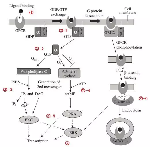
G protein-coupled receptors are one of the major targets for drug discovery, 460 of 1286 drugs approved by the FDA for approval in 2016 are targets of G protein-coupled receptors, accounting for more than one-third of the total. The leading supplier of instrumentation reagents provides a functional assessment of each stage of the cellular physiological response to this target. Physiologic response processes activated by G-protein coupled receptors can be used for each of the key biomarkers for functional assessment. The intracellular partial configuration of the G protein-coupled receptor is changed so that its binding to the G protein will be enhanced after the G-protein coupled receptor is activated by the candidate drug. After binding of the G protein and the G-protein coupled receptor, G ɑ-subunit activity changes so that the G ɑ-subunit more easily binds to GTP and dissociates from both subunits. After G ɑ-subunit and GTP are bound, the cascade of downstream signals is activated according to the different changes of their types and the activity of different effector proteins, which will result in changes in the concentration of the second messenger and in the concentration of the second messenger to activate subsequent transcription factor pathways, as well as the activation of transcription factors to regulate cellular protein expression, and cytological responses. G-protein-coupled receptors, upon activation, are phosphorylated and the β-arrestin and phosphorylated G-protein coupled receptors mediate their internalization upon binding. The internalized G-protein coupled receptors are returned to the surface of cells or into the lysosome degradation, which is the antagonist pathway of G-protein coupled receptor after activation. The mainstream detection methods will be summarized as follows:
Classification of cell functional assessment methods
According to the single physiological reaction process, the cell functional assays can be divided into the following four categories:
1.Determining the initial alteration of the target protein by the candidate drug, the most common is the binding of a candidate drug to a target receptor located on the cell membrane or the binding of a target receptor to a natural ligand, and commonly used assays that include cell-based ELISA and flow cytometry can be used to determine candidate drug-receptor conjugates. Differ from the target receptor recombination purified affinity evaluation, the target receptor on the cell membrane is closer to the true physiological conditions, the other receptors and the effector protein on the cell may be involved in the activation of the target protein changes, and including more reference value. Moreover, many membrane proteins are multi-permeable membrane structures. It is difficult to purify the receptor proteins that have a complete tertiary structure conformation (eg, G-protein coupled receptors are membrane-permeable).
2.Determination on the concentration of key biomarkers in signaling pathways during signal transduction, such as cAMP, DAG, IP3 (TR-FRET, ELISA) and Ca+2 (FLIPR) during GPCR activation; key effect proteins phosphorylation and other modified concentrations (TR-FRET, WB); concentration determination of key enzyme; kinetic transport process (internalization) etc.
3.Determination of the level of transcription and translation that cause cellular responses. For example, RT-qPCR was used to measure the level of transcription RNA. The transcription factor activity was measured by inserting a foreign reporter gene behind the promoter.
4. Cell proliferation or death was measured by MTT, CCK-8, Alarmable, ATP assay, LDH assay, proteomic or genomic changes of the cells (flow cytometry cycles of apoptosis), cell migration and migration capacity changes (scratch, invasion, transwell experiments), determination of cell exudates such as TNF and IL quantification of PBMC cells (ELISA, TR-FRET).
All the mentioned above is the brief summary of cell function evaluation methods, from which we can lean that there are various methods can be applied to evaluate cells function, but how to choose the appropriate test method to satisfy the research purpose still is a key point. Researchers already shared several suggestions on choosing an appropriate method:
1.In the initial stage of basic research and target identification of drug development. Scientists should combine with the literature and process basic functional and functional experiments at various stages of the cell's single physiological response process, that is signal reception (target protein binding), signal amplification (second messenger or kinase assay), transcriptional translation (RT-qPCR or reporter gene system), final response (cell viability assay, secretion assay), establishing a complete network of cell responses after drug stimulation and assessing the effectiveness of the target.
2.In the drug screening phase, initial high-throughput screening of drugs is required based on experimental ease-of-use, protein-binding assays (category 1) or cell viability assays (category 4). The most important part of the functional assessment stage is the assessment of the level of binding (Class 1) and the final response level (Class 4) of candidate drugs of relatively pure activity and the accurate data presentation to be established in connection with the results of animal experiments.
3.In the quality control phase, the stability and precision of the method should be considered, binding force assessment of purified protein levels (category 4) and methods of cell functional assessment (categories 2, 3 and 4) will be needed for release.
Functional assessment of G protein-coupled receptors and ADCC effects
G protein-coupled receptors are one of the major targets for drug discovery, 460 of 1286 drugs approved by the FDA for approval in 2016 are targets of G protein-coupled receptors, accounting for more than one-third of the total. The leading supplier of instrumentation reagents provides a functional assessment of each stage of the cellular physiological response to this target. Physiologic response processes activated by G-protein coupled receptors can be used for each of the key biomarkers for functional assessment. The intracellular partial configuration of the G protein-coupled receptor is changed so that its binding to the G protein will be enhanced after the G-protein coupled receptor is activated by the candidate drug. After binding of the G protein and the G-protein coupled receptor, G ɑ-subunit activity changes so that the G ɑ-subunit more easily binds to GTP and dissociates from both subunits. After G ɑ-subunit and GTP are bound, the cascade of downstream signals is activated according to the different changes of their types and the activity of different effector proteins, which will result in changes in the concentration of the second messenger and in the concentration of the second messenger to activate subsequent transcription factor pathways, as well as the activation of transcription factors to regulate cellular protein expression, and cytological responses. G-protein-coupled receptors, upon activation, are phosphorylated and the β-arrestin and phosphorylated G-protein coupled receptors mediate their internalization upon binding. The internalized G-protein coupled receptors are returned to the surface of cells or into the lysosome degradation, which is the antagonist pathway of G-protein coupled receptor after activation. The mainstream detection methods will be summarized as follows:
 1.Combination of Ligand and receptor, cell-level affinity assessment experiments.
In the practical application process, especially in the early stage of the method development, the evaluation method which takes the protein in cell surface as target usually adopt Cell-Basd ELISA or flow cytometry, and the mode can be varied:
. Direct detection: Candidate drugs are directly detected by fluorescent labeling
. Indirect detection: Fluorescent / enzyme-labeled secondary antibody against non-target drug binding sites (eg, Fc-terminal rabbit anti-human IgG1 conjugated HRP as secondary antibody )
. Competition assays: labeling the native ligands for the target protein with competing drug candidates
In general, indirect assay and competition assay are more common and comparable, suitable for screening multiple candidate drugs at the same time. It should be noted that competition testing is directed against a single binding epitope affinity assessment of the candidate drug and the target protein. For direct and indirect methods, however, evaluate the affinity of candidate drugs and the entire structure of the target protein.
2.Ca2+, IP1, cAMP and other second messengers detection
Ca2+ and fluorescent dyes (Fluo-3AMA, etc.) combined with the emission of light generated after excited by a specific wavelength, the intensity of the emitted light is linearly related to the concentration of calcium ions. Since the change of Ca2+ concentration after stimulation is a transient process, it will increase and return to the baseline within a short time. The detection of Ca2+ is a real-time dynamic process. During the whole experiment, the competition of sample and test need to control in a very short period of time so that fluorescence Imaging Plate Reader FLIPR ™ technology and instruments for instantaneous ion concentration change detection instrument will be practical technique to study ion channel and GPCR functions, as it is featured with the automated injection, microplate mode, real-time dynamic detection process, have the ability to monitor agonist, potentiator and antagonist effects of high-throughput, candidate drugs in a single experiment by controlling the loading sequence and concentration of candidate and natural ligands.
1.Combination of Ligand and receptor, cell-level affinity assessment experiments.
In the practical application process, especially in the early stage of the method development, the evaluation method which takes the protein in cell surface as target usually adopt Cell-Basd ELISA or flow cytometry, and the mode can be varied:
. Direct detection: Candidate drugs are directly detected by fluorescent labeling
. Indirect detection: Fluorescent / enzyme-labeled secondary antibody against non-target drug binding sites (eg, Fc-terminal rabbit anti-human IgG1 conjugated HRP as secondary antibody )
. Competition assays: labeling the native ligands for the target protein with competing drug candidates
In general, indirect assay and competition assay are more common and comparable, suitable for screening multiple candidate drugs at the same time. It should be noted that competition testing is directed against a single binding epitope affinity assessment of the candidate drug and the target protein. For direct and indirect methods, however, evaluate the affinity of candidate drugs and the entire structure of the target protein.
2.Ca2+, IP1, cAMP and other second messengers detection
Ca2+ and fluorescent dyes (Fluo-3AMA, etc.) combined with the emission of light generated after excited by a specific wavelength, the intensity of the emitted light is linearly related to the concentration of calcium ions. Since the change of Ca2+ concentration after stimulation is a transient process, it will increase and return to the baseline within a short time. The detection of Ca2+ is a real-time dynamic process. During the whole experiment, the competition of sample and test need to control in a very short period of time so that fluorescence Imaging Plate Reader FLIPR ™ technology and instruments for instantaneous ion concentration change detection instrument will be practical technique to study ion channel and GPCR functions, as it is featured with the automated injection, microplate mode, real-time dynamic detection process, have the ability to monitor agonist, potentiator and antagonist effects of high-throughput, candidate drugs in a single experiment by controlling the loading sequence and concentration of candidate and natural ligands.
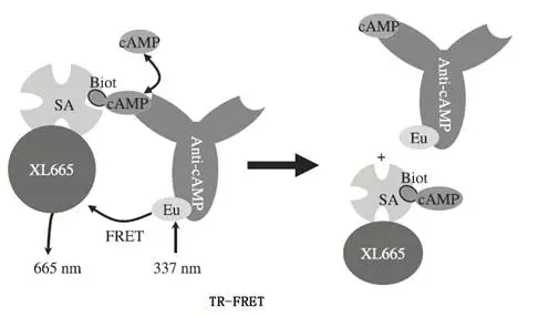
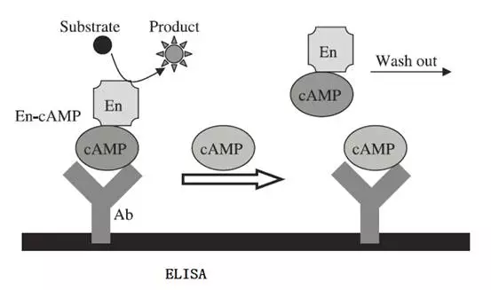
 Second messengers such as IP1, cAMP and other small molecules can generally be detected quantitatively by the competitive method such as ELISA, TR-FRET, polarized fluorescence etc. There are several suppliers in the market that provide relevant kits for testing. It should be noted that IP1, cAMP standards need to be added before testing to establish a standard curve to determine the linear detection range of the kit and optimize the number of cells added.
 3.Detection of phosphorylated ERK
For phosphorylated ERK and other key macromolecular protein activation can be detected with AlphaScreen technology, the two microspheres tested captured the ERK protein and the phosphorylation site respectively, energized to donate energy through donor microspheres, and acceptor microspheres that are physically covalently attached are stimulated to emit light upon receiving energy The method can avoid the situation that the energy transfer will be greatly attenuated when the TR-FRET technique performs the quantitative determination of macromolecules, and obtain better signal-to-noise ratio.
3.Detection of phosphorylated ERK
For phosphorylated ERK and other key macromolecular protein activation can be detected with AlphaScreen technology, the two microspheres tested captured the ERK protein and the phosphorylation site respectively, energized to donate energy through donor microspheres, and acceptor microspheres that are physically covalently attached are stimulated to emit light upon receiving energy The method can avoid the situation that the energy transfer will be greatly attenuated when the TR-FRET technique performs the quantitative determination of macromolecules, and obtain better signal-to-noise ratio.
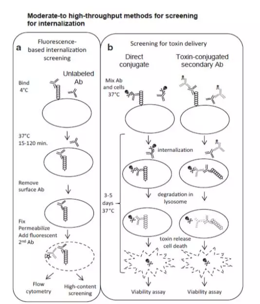
 4.Internalization of G-protein coupled receptors
Conventional internalization method is to fluorescently label the ligand of the receptor, and after internalization of the cell binding, the fluorescent-labeled ligand bound to the cell surface is removed by pickling or enzymatic digestion, and the intensity of the fluorescent label is detected to represent the strength of internalization or the ligand may be labeled with a toxic molecule. Recipients and ligands internalize into the cell and release toxic molecules, which can be used to determine the intensity of internalization by measuring cell activity. But these two methods are relatively poor in operability or stability. At present, pH-sensitive fluorescent dyes can avoid the pickling process, ensuring that fluorescent dyes that do not enter the cell have no fluorescent signal in extracellular neutral or weakly alkaline medium, and signals are generated in the acidic environment after internalization into the cells. Pickling to avoid real-time dynamic internalization of the method of detection.
4.Internalization of G-protein coupled receptors
Conventional internalization method is to fluorescently label the ligand of the receptor, and after internalization of the cell binding, the fluorescent-labeled ligand bound to the cell surface is removed by pickling or enzymatic digestion, and the intensity of the fluorescent label is detected to represent the strength of internalization or the ligand may be labeled with a toxic molecule. Recipients and ligands internalize into the cell and release toxic molecules, which can be used to determine the intensity of internalization by measuring cell activity. But these two methods are relatively poor in operability or stability. At present, pH-sensitive fluorescent dyes can avoid the pickling process, ensuring that fluorescent dyes that do not enter the cell have no fluorescent signal in extracellular neutral or weakly alkaline medium, and signals are generated in the acidic environment after internalization into the cells. Pickling to avoid real-time dynamic internalization of the method of detection.
 5.ADCC detection by gene reporter method and ADCC detection by PBMC method
PBMC method for the detection of ADCC effect is the use of fresh peripheral blood mononuclear cells (PBMC) as effector cells, detection and target cells of the antibody, mediated by effector cells killing target cells, commonly used LDH as biomarkers of target cell death to process quantitative detection. In essence, the gene reporter method is an antibody that interacts with target cells, activating the effector cell signaling pathway and the transcription factor expression reporter protease, and characterizes the ADCC activity of the antibody by quantifying the reporter protease. From the reality of the method, PBMC is more convincing from a methodological point of view and there must be such experimental data during the final validation phase of antibody ADCC function. However, the PBMC method also has its drawbacks. The fresh source of peripheral blood makes the material preparation of the method costly and complicated. The complexity and lot differences of fresh blood also make the method less stable and precise. After confirming the equivalency of the gene reporter method and the PBMC method, the gene reporter method would be a better choice, which has better stability and is suitable for function evaluation and quality release.
5.ADCC detection by gene reporter method and ADCC detection by PBMC method
PBMC method for the detection of ADCC effect is the use of fresh peripheral blood mononuclear cells (PBMC) as effector cells, detection and target cells of the antibody, mediated by effector cells killing target cells, commonly used LDH as biomarkers of target cell death to process quantitative detection. In essence, the gene reporter method is an antibody that interacts with target cells, activating the effector cell signaling pathway and the transcription factor expression reporter protease, and characterizes the ADCC activity of the antibody by quantifying the reporter protease. From the reality of the method, PBMC is more convincing from a methodological point of view and there must be such experimental data during the final validation phase of antibody ADCC function. However, the PBMC method also has its drawbacks. The fresh source of peripheral blood makes the material preparation of the method costly and complicated. The complexity and lot differences of fresh blood also make the method less stable and precise. After confirming the equivalency of the gene reporter method and the PBMC method, the gene reporter method would be a better choice, which has better stability and is suitable for function evaluation and quality release.

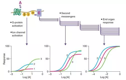
 Association of various detection methods
Existing research data show that there is a certain correlation between the various detection methods, but not absolute relevance, even with the same drug candidate in different cells may also have different dose-response curve:
The essence of functional method development
The process of cellular physiological response is a complex and continuous process, the signal transduction and amplification of the signal in the cell is continuous and multifactorial, non-linear process from the initial cell signal to the final cell response. The experimental results provided by each detection method are fragmented information, and the functional evaluation method can display and collect the fragmented information, but not able to make a positive judgment on the pros and cons of the candidate drugs. However, it can provide a certain degree of correlation for the follow-up zoological experiments and even clinical experiments to summarize the experience. In the whole information collection process, the authenticity of the data is very important, the accuracy and authenticity of the functional evaluation method need to be interpreted by the experimental method itself.
Review of the above functional experiments, all of the functional evaluation of the essence is about affinity determination and biomarker concentration determination. Affinity determination is the basis, however, only the combination can make it functional. The signals of the cells caused by the binding are multiplied and amplified, and the exact quantification of each biomarker in this process is the key to functional evaluation regardless of the detection technique. Quantitation of Ca2+, IP1, cAMP, phosphorylation of ERK, the quantification of firefly enzyme, the number of viable/dead cells, and the signal and biomarker concentrations should be linearly correlated during quantitation. This covert precondition is often neglected and become one of the key factors that result in experimental data of functional assessment methods were misinterpreted and have poor stability. The non-linear transmission of intracellular signals caused by the binding of candidate drugs is an objective response to the process of cellular physiological responses. It is a mistake to detect the nonlinear relationship between signals and biomarkers.
Association of various detection methods
Existing research data show that there is a certain correlation between the various detection methods, but not absolute relevance, even with the same drug candidate in different cells may also have different dose-response curve:
The essence of functional method development
The process of cellular physiological response is a complex and continuous process, the signal transduction and amplification of the signal in the cell is continuous and multifactorial, non-linear process from the initial cell signal to the final cell response. The experimental results provided by each detection method are fragmented information, and the functional evaluation method can display and collect the fragmented information, but not able to make a positive judgment on the pros and cons of the candidate drugs. However, it can provide a certain degree of correlation for the follow-up zoological experiments and even clinical experiments to summarize the experience. In the whole information collection process, the authenticity of the data is very important, the accuracy and authenticity of the functional evaluation method need to be interpreted by the experimental method itself.
Review of the above functional experiments, all of the functional evaluation of the essence is about affinity determination and biomarker concentration determination. Affinity determination is the basis, however, only the combination can make it functional. The signals of the cells caused by the binding are multiplied and amplified, and the exact quantification of each biomarker in this process is the key to functional evaluation regardless of the detection technique. Quantitation of Ca2+, IP1, cAMP, phosphorylation of ERK, the quantification of firefly enzyme, the number of viable/dead cells, and the signal and biomarker concentrations should be linearly correlated during quantitation. This covert precondition is often neglected and become one of the key factors that result in experimental data of functional assessment methods were misinterpreted and have poor stability. The non-linear transmission of intracellular signals caused by the binding of candidate drugs is an objective response to the process of cellular physiological responses. It is a mistake to detect the nonlinear relationship between signals and biomarkers.
Your email address will not be published. Required fields are marked *