Overview of T Cell Activation and Expansion
In recent years, the boom in immune cell therapy has led to an increased interest in the industrial production of T cells. To accomplish successful cell therapy, a large number of CAR-T or other types of T cells are required. Cell activation technology, cytokine dosage, and easy-to-use bioreactors are key factors in T cell production.
Signals for T-cell Activation
 Fig. 1
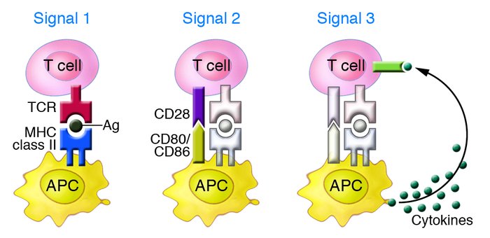
Three signals are required for antigen-specific T-cell activation. (Gutcher I and Becher B., 2007)
- Signal 1. The interaction of antigen-specific T cell receptors (TCR/CD3) on T cells with antigen peptide-MHC molecular complexes on antigen-presenting cells (APCs), i.e., the process of antigen recognition. In addition, CD4 and CD8 molecules on the surface of T cells act as co-receptors that bind to MHC-II and MHC-I class I molecules, respectively.
- Signal 2. Co-stimulatory signals - provided by interactions between co-stimulatory molecules on the APC and the corresponding receptor molecules on the surface of the T cell. Of these, the most important is the binding of CD28 molecules on the surface of T cells to the corresponding ligands on the surface of APCs, B7-1 (CD80) and B7-2 (CD86). Notably, the CTLA-4 receptor expressed by activated T cells also binds B7-1 and B7-2 but transmits T cell inhibitory signals.
- Signal 3. Cell-secreted cytokines, such as IL-2, promote T cell clonal expansion and the formation of daughter cells that differentiate into effector and memory T cells.
Theoretical Basis of T Cell Expansion and Culture in Vitro
According to the three stages of T cell activation, we can simulate the three signals of T cell activation through different stimulators to achieve T cell expansion and culture in vitro.
-
Modeling of the first signal
CD3 antibody is usually used, which triggers intracellular ITAM signaling upon binding to TCR/CD3 on the surface of T cells. The signaling pathway downstream of TCR/CD3 can be activated through Lck/ZAP-70 transduction. -
Modeling of the second signal
The most commonly used is the CD28 antibody. By binding to CD28 on the surface of T cells, it can promote the proliferation and activation of T cells. -
Modeling of the third signal
T cells are often stimulated in culture by the use of IL-2, which is added to the culture medium.IL-2 is the most important cytokine for stimulating T-cell proliferation. It promotes T cell activation and proliferation by specifically binding to the IL-2 receptor on the surface of T cells. Resting T cells basically do not express IL-2R and do not receive IL-2 stimulation; only when T cells are activated can they express IL-2R and receive the stimulatory effect of IL-2.
T Cell Expansion and Culture
In vitro, T cells are stimulated with a combination of CD3 and CD28 antibodies to mimic the dual signals of T cell activation in vivo. It is currently the most widely used method for T cell activation and expansion application.
-
Antibody-based method
The experimental steps of the antibody method for the expansion of cultured T cells include antibody encapsulation, closure, isolation of lymphocytes, and cell stimulation and culture. -
Magnetic bead method
Magnetic beads are typically 4-5 μm in size, which is similar to the size of cells. By coupling CD3/CD28 to the magnetic beads, the process of activating T-cells in vivo by antigen-presenting cells can be simulated. -
Multimer method
In CD3/CD28 coupled multimers, CD3 and CD28 form a complex with a soluble multimer. This complex binds to T cells and promotes their activation. The addition of D-Biotin causes the complex to dissociate, aborting the stimulatory signal to the T cells. Thus, the duration of T-cell activation can be precisely regulated using this method.
Creative Bioarray Relevant Recommendations
Creative Bioarray offers a wide variety of human T cells, including Human T Cells-Negatively Selected (HTC), Normal PB Pan T Cells, Normal PB CD8+/CD45RA+ Naïve Cytotoxic T Cells, Normal PB CD8+ T Cells, Normal PB CD4+/CD45RO+ Memory T Cells, Normal PB CD4+ Helper T Cells, etc. These cells can be used for T-cell research applications.
Reference
- Gutcher I, Becher B. (2007). "APC-derived cytokines and T cell polarization in autoimmune inflammation." J Clin Invest. 117 (5), 1119-27.

Your email address will not be published. Required fields are marked *