
4T1: The Syngeneic "Gold Standard" for Triple-Negative Breast Cancer Metastasis Research
When preclinical researchers need a model that recapitulates the full malignant arc of human breast cancer—from primary tumor formation to spontaneous multi-organ metastasis—4T1 remains the undisputed benchmark. Isolated in 1974 from a spontaneous mammary carcinoma in a BALB/c mouse, this cell line is far more than a convenient in vitro tool. It is the most widely validated syngeneic, immunocompetent model for stage IV triple-negative breast cancer (TNBC), enabling studies that xenograft platforms simply cannot support.
Origin & the Syngeneic Advantage
The 4T1 cell line was originally derived from a spontaneously arising mammary tumor in a BALB/c mouse and was subsequently selected for resistance to 6-thioguanine.
Unlike human tumor xenografts that require immunodeficient hosts, 4T1 is transplanted into syngeneic (allograft) BALB/c mice with fully intact immune systems.
This immunological match eliminates graft rejection and unlocks a critical experimental dimension: the ability to study tumor–immune crosstalk, including T-cell exhaustion, myeloid-derived suppressor cell (MDSC) recruitment, and resistance to immune-checkpoint blockade—phenomena that define clinical TNBC but are invisible in immunocompromised models.
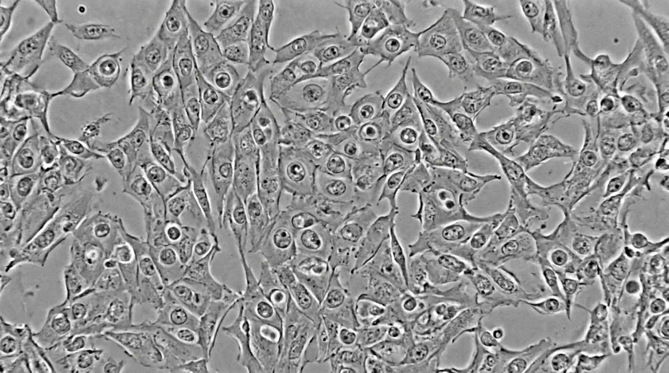
Four Hallmarks That Make 4T1 Indispensable
1. Triple-Negative Phenotype
4T1 cells lack expression of the estrogen receptor (ER), progesterone receptor (PR), and HER2/neu. This ER-/PR-/HER2- profile mirrors the most aggressive human breast cancer subtype, which carries the worst prognosis and the fewest targeted therapeutic options.
2. Potent Tumorigenicity
Even at low inoculum densities, 4T1 cells reliably engraft and form palpable primary tumors. When implanted orthotopically into the mammary fat pad, tumors progress rapidly, providing a compressed timeline for therapeutic intervention studies.
3. Spontaneous, Multi-Organ Metastasis
The defining feature of 4T1 is its propensity to metastasize from the primary site to distant organs in a pattern that closely parallels human breast cancer. Following orthotopic implantation, tumor cells disseminate hematogenously to the lungs, liver, bone, and brain—the same organs most commonly affected in advanced human disease.
4. Immunosuppressive Microenvironment
4T1 tumors are notoriously "cold." They recruit large populations of MDSCs and regulatory T cells that suppress anti-tumor immunity, making the model a stringent stress-test for immunotherapy candidates. Agents that show efficacy in 4T1 are often viewed as having high clinical translation potential precisely because they overcome such a hostile microenvironment.
Primary Research Applications
Metastatic Cascade Dissection
4T1 enables stepwise investigation of the metastatic sequence: local invasion, intravasation, survival in circulation, extravasation, and colonization of secondary organs. It has been instrumental in validating the pre-metastatic niche hypothesis—the concept that primary tumors remotely precondition distant organs to support future metastatic seeding.
Immuno-Oncology & Therapy Development
Because the model retains a functional immune system, it is the platform of choice for evaluating:
- Immune-checkpoint inhibitors (e.g., anti-PD-1, anti-CTLA-4)
- Cancer vaccines and oncolytic virotherapy
- CAR-T and CAR-NK cell therapies
- Combination regimens targeting both tumor cells and immunosuppressive stroma
Anti-Metastatic Drug Screening
Given its reliable distal metastasis, 4T1 is the gold-standard in vivo filter for compounds designed to prevent or treat metastatic disease rather than simply shrink primary tumors.
Tumor Microenvironment & Imaging Studies
4T1 is easily engineered to express luciferase, GFP, or RFP, permitting non-invasive, real-time monitoring of tumor growth and metastatic burden via bioluminescence or fluorescence imaging.
Related Models for Comparative Biology
The 4T1 family includes several derivative lines that serve as powerful genetic and phenotypic controls:
| Cell Line | Origin / Relationship | Metastatic Potential | Research Utility |
|---|---|---|---|
| 4T1 | Parental line from BALB/c mammary tumor | High (lung, liver, bone, brain) | General metastasis & immunotherapy |
| 67NR | Derived from the same primary tumor as 4T1 | Non-metastatic (grows locally only) | Identifying genes required for dissemination |
| 168FARN | Related subline | Weak metastasis | Intermediate metastatic phenotyping |
| 66cl4 | Related subline | Lung-specific metastasis | Organotropism studies |
By comparing 4T1 with 67NR and other sublines, researchers can perform differential gene expression and functional screens to isolate the molecular drivers of metastasis—an approach that has yielded numerous mechanistic insights into EMT, anoikis resistance, and organ-specific colonization.
Experimental Considerations
Implantation Route Dictates Metastatic Pattern
While orthotopic mammary fat pad injection most closely mimics spontaneous human breast cancer metastasis, experimental metastasis models (e.g., tail vein injection) bias toward pulmonary colonization and are useful for isolating specific steps in the cascade.
Proliferation vs. Metastasis Controls
To distinguish anti-proliferative effects from genuine anti-metastatic activity, primary tumors can be surgically excised at a standardized volume, with subsequent monitoring of metastatic outgrowth in target organs.
Timeline & Endpoints
Orthotopic 4T1 models typically develop measurable primary tumors within 1–2 weeks, with macroscopic lung metastases evident by 3 weeks. Bioluminescent imaging allows earlier detection of micrometastatic seeding.
Conclusion
The 4T1 model occupies a unique position in preclinical oncology: it is simultaneously aggressive, metastatic, triple-negative, and immunologically intact. No single xenograft or humanized platform can replicate this combination of features. Its ability to mirror the metastatic organotropism and immune evasion strategies of human TNBC makes it the ultimate proving ground for therapies aimed at late-stage disease. Any compound or biologic that demonstrates efficacy against 4T1 metastasis carries a strong rationale for clinical advancement.
Source Your 4T1 Models & Metastasis Assays from Creative Bioarray
Whether you need authenticated cell stocks, luciferase-enabled reporter lines, or fully managed in vivo efficacy studies, Creative Bioarray provides end-to-end solutions for 4T1-based research:
Cell Lines & Reporter Models
- Wild-type 4T1 cells validated for high tumorigenicity and spontaneous metastasis
- 4T1-Luc2 stable cell line for quantitative bioluminescence imaging of metastatic burden
- 4T1-Epcam stable cell line for studies targeting epithelial adhesion molecules
- COL1A1 knockout 4T1 cell line for microenvironment and extracellular matrix research
In Vivo Model Services
- Syngeneic tumor models in immunocompetent BALB/c mice, benchmarked against clinical immune-checkpoint inhibitors
- Orthotopic breast cancer models implanting 4T1 into the mammary fat pad to recapitulate spontaneous metastasis
- Metastatic model services with ex vivo organ analysis, histopathology, and quantitative imaging endpoints
In Vitro Migration & Invasion Assays
- Cell migration and invasion assay services including Transwell and 3D spheroid invasion platforms to deconvolute metastatic mechanisms in vitro before advancing to in vivo studies
From cell-line authentication and mycoplasma testing to GLP-compliant in vivo pharmacology, Creative Bioarray's integrated platform reduces experimental variability and accelerates the path from mechanistic hypothesis to publication-ready data. Contact their team to customize your 4T1 study design today.
Your email address will not be published. Required fields are marked *