
HeLa Cells: Why This Classic Cell Line Still Matters
From visualizing cell division to investigating gene function, HeLa cells remain a widely used model in biomedical research. Their capacity for sustained growth and compatibility with many experimental workflows make them useful for exploring how human cells function—and how those functions become disrupted in cancer.
However, familiarity can encourage assumptions. HeLa cells are cancer-derived, carry an abnormal genome, and can differ substantially between laboratory stocks.
Understanding these characteristics is essential to using HeLa cells effectively and interpreting their results accurately.
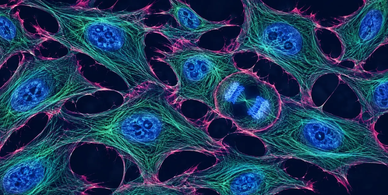
What Are HeLa Cells?
HeLa is a human cervical cancer cell line derived from tissue collected from Henrietta Lacks in 1951. Its name combines the first two letters of her first and last names. The cell line’s history is inseparable from her contribution to biomedical research.
HeLa cells have epithelial-like morphology and are commonly maintained as adherent cultures. They contain human papillomavirus type 18, or HPV18, sequences and have an aneuploid genome—meaning their chromosome complement differs from that of normal diploid human cells.
They are often described as “immortal.” In this context, immortality refers to a population’s capacity for continued proliferation under suitable culture conditions.
| Characteristic | HeLa cell profile | Implication for research |
|---|---|---|
| Species | Human | Useful for investigating human cellular processes |
| Tissue origin | Cervix | Provides a cervical cancer-derived experimental context |
| Morphology | Epithelial-like | Compatible with many microscopy workflows |
| Common growth format | Adherent monolayer | Convenient for imaging on suitable culture surfaces |
| Viral background | HPV18-associated | Relevant when interpreting growth control and stress responses |
| Genome | Aneuploid | Gene dosage and DNA-content measurements require careful interpretation |
| Population identity | Multiple sublines and laboratory variants | Exact stock information matters for reproducibility |
Why Are HeLa Cells So Widely Used?
Sustained Growth Supports Repeated Experiments
HeLa cells can be expanded to support repeated experiments, assay development, and the establishment of working cell banks. This makes them practical when researchers need sufficient material for parallel imaging, molecular analysis, and functional assays.
Their durability is a major advantage, but it does not remove the need to control cell density, passage history, and culture conditions.
Adherent Growth Makes Cellular Structures Accessible to Imaging
In suitable cultures, individual cells provide visible nuclear and cytoplasmic regions that can be examined by fluorescence microscopy.
This supports experiments investigating:
- Protein localization and redistribution.
- Cytoskeletal organization.
- Organelle morphology.
- Chromosome behavior during division.
- Responses to genetic or chemical perturbations.
The value of HeLa for imaging is demonstrated by large-scale studies that combined gene silencing with time-lapse microscopy to identify genes involved in cell division.
A Broad Experimental History Helps Researchers Develop Assays
Published HeLa studies provide starting points for selecting markers, evaluating experimental designs, and comparing phenotypes.
However, a published protocol should be treated as a starting framework. Its performance still needs to be confirmed in the specific stock, culture conditions, and instrument configuration being used.
Key Applications of HeLa Cells
1. Immunofluorescence and Subcellular Localization
HeLa cells are useful for studying where proteins are located and whether their distribution changes after treatment.
A fixed-cell experiment might combine a target-specific antibody with a cytoplasmic marker and a nuclear counterstain. This can help distinguish nuclear accumulation, cytoplasmic redistribution, or changes in organelle-associated patterns.
For convincing results, validate antibody specificity, optimize fixation and permeabilization, and include appropriate controls. A visually attractive image alone does not establish that the detected signal represents the intended protein.
2. Cell Division and Functional Genomics
HeLa cells support studies linking gene function to observable phenotypes.
For example, fluorescent chromatin reporters can reveal changes in chromosome alignment, segregation, and the timing of division. Published genome-wide RNA interference work used fluorescently labeled HeLa chromosomes and automated imaging to investigate these processes.
Genetic perturbation experiments should account for off-target effects and, where feasible, include independent targeting reagents or rescue experiments.
3. Cancer Biology and HPV-Associated Mechanisms
HeLa cells provide a model for investigating aspects of HPV-associated cancer biology.
Their viral oncogene background is particularly relevant to growth regulation. Experiments repressing HPV oncogenes in HeLa cells have demonstrated reactivation of p53- and retinoblastoma-associated pathways, with effects on proliferation, senescence, and apoptosis.
Consequently, HeLa should not be treated as a model with normal tumor suppressor regulation. The pathway being studied must be considered alongside the cell line’s biological background.
4. Compound Response and Cell Death Studies
HeLa cells can be used to examine changes in proliferation, morphology, and cell survival following compound exposure.
Interpretation improves when the readouts address distinct biological questions. For instance, a reduction in a metabolic assay signal may reflect fewer cells, altered metabolism, or cell death. Additional measurements help distinguish these possibilities.
Likewise, condensed nuclei can support a cell death assessment, but nuclear morphology alone does not establish apoptosis.
The Limitations Researchers Should Understand
HeLa Cells Do Not Represent Normal Cervical Tissue
HeLa is a cancer-derived model. Its behavior cannot be assumed to reproduce normal epithelial physiology, intact tissue organization, or the diversity of cervical tumors.
For conclusions involving tissue-specific function or therapeutic relevance, consider validation in additional models, such as other relevant cancer cell lines, primary cells, or appropriately characterized organoids.
Different HeLa Stocks Can Produce Different Results
“HeLa” does not describe a single experimentally identical population across laboratories.
A multi-omics study examining stocks from 13 laboratories found substantial molecular and phenotypic differences between HeLa variants, as well as progressive divergence during extended passaging.
Record the supplier, accession or catalog number, subline, passage history, and any introduced modifications. These details help explain differences that might otherwise be attributed entirely to experimental technique.
Morphology Cannot Confirm Identity
HeLa has a well-documented history of cross-contaminating other cultures. A healthy-looking epithelial monolayer is therefore insufficient evidence that a culture has the expected identity.
Short tandem repeat, or STR, profiling supports human cell line identity verification. Mycoplasma testing addresses a separate quality concern; one test does not replace the other.
Practical Tips for Reproducible HeLa Experiments
Follow the instructions for the exact stock. Media, serum requirements, and growth formats may differ between parental cultures and derivatives. Avoid assuming that one HeLa protocol applies to every variant.
Standardize the starting conditions. Match seeding density, attachment time, and treatment timing across comparison groups. Check the actual culture rather than relying solely on elapsed time.
Use a documented passage window. Establish working banks and limit unnecessary continuous culture. Record passage numbers consistently.
Confirm identity and culture quality. Include authentication and mycoplasma testing in the laboratory’s quality management workflow.
Choose measurements that answer the question. Combine imaging with complementary molecular or functional assays when a single readout cannot distinguish competing explanations.
Keep image acquisition comparable. Avoid saturated signals, retain original data, and apply consistent analysis methods across groups.
Support Reliable Cell-Based Research With Creative Bioarray
Reliable experiments begin with well-characterized cells. Whether you are establishing a HeLa-based assay or investigating unexpected changes in an existing culture, confirming cell identity and culture quality provides a stronger foundation for interpreting results.
Creative Bioarray offers Cell Line Testing and Quality Control Services, including STR-based cell line authentication and mycoplasma detection services. Explore the available options to support your cell culture quality control workflow and discuss the testing appropriate for your research material.
Your email address will not be published. Required fields are marked *