
Beyond DAPI: Are You Using the Right Nuclear Stain for Your Experiment?
Fix, permeabilize, incubate with antibodies, and finish with DAPI.
For many researchers, nuclear counterstaining is almost automatic. The familiar blue nuclei provide a convenient reference for cell counting, tissue organization, and protein localization.
But a reliable routine stain is not necessarily the right stain for every experiment.
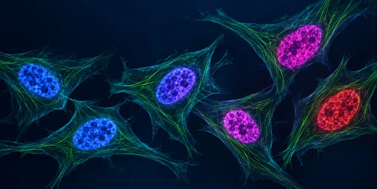
Why DAPI Remains a Popular Nuclear Counterstain?
DAPI, short for 4’,6-diamidino-2-phenylindole, preferentially associates with A–T-rich regions in the minor groove of double-stranded DNA. DNA-bound DAPI produces bright blue fluorescence, with approximate excitation and emission maxima of 358 and 461 nm, respectively.
For routine fixed-cell immunofluorescence, its advantages are straightforward:
- Clear nuclear contrast for locating and segmenting nuclei.
- Convenient integration into many established staining workflows.
- Blue emission that often fits alongside green and red antibody labels.
- Useful visualization of nuclear size, shape, and chromatin organization.
If the sample is fixed, the blue channel is available, and nuclear identification is the main goal, DAPI remains a practical choice.
Problems arise when its suitability is assumed without considering the sample, imaging platform, or intended measurement.
Pitfall 1: Treating DAPI as a Universal Live-Cell Nuclear Stain
Cell permeability changes what the signal means
DAPI generally enters intact living cells less readily than Hoechst 33342. Its uptake in live samples can therefore be weak or inconsistent, depending on the cell type and staining conditions. Hoechst 33342 is more readily cell-permeant and is commonly used to label nuclei in both living and fixed cells.
This distinction matters when interpreting a live sample.
Under a validated dye-exclusion protocol, strong DAPI uptake can indicate compromised plasma membrane integrity. However, DAPI permeability is not an absolute on/off property: a positive nucleus should be interpreted in the context of concentration, incubation time, cell type, and appropriate controls.
A weak DAPI signal in a live sample may also reflect poor dye entry rather than an absence of cells.
Hoechst 33342 is an option, not a guarantee of biological neutrality
For short live-cell observations, Hoechst 33342 may provide more consistent nuclear labeling. Nevertheless, DNA binding and repeated short-wavelength illumination can perturb the cells being observed.
A useful stain should provide sufficient contrast while preserving the biological behavior relevant to the experiment. That requires testing the complete staining and imaging workflow.
Pitfall 2: Adding DAPI Before Planning the Fluorescence Panel
A blue nucleus may appear visually distinct from green or red labels, but display colors do not determine spectral compatibility.
Before selecting a nuclear stain, check:
- Available excitation sources and detector ranges.
- Excitation and emission spectra of every fluorophore.
- Potential excitation of multiple labels by the same light source.
- Emission bleed-through between detection channels.
- Sample autofluorescence and the relative brightness of each target.
DAPI and Hoechst also occupy a similar spectral region. Replacing DAPI with Hoechst 33342 may improve live-cell uptake, but it generally does not free the blue detection channel.
When a far-red nuclear stain may help
DRAQ5 is a cell-permeant, far-red DNA stain used with living and fixed samples. It can be useful when a nuclear reference is needed outside the blue channel.
However, moving the nuclear signal into the far-red region also uses spectral space that may already be assigned to an antibody or reporter. For example, potential overlap with Cy5- or Alexa Fluor 647-based detection must be evaluated.
Single-stain controls acquired using the intended imaging settings help reveal signal leakage that may be difficult to recognize in merged images.
Pitfall 3: Confusing Nuclear Labeling with Cell Viability
A fluorescent nucleus does not, by itself, demonstrate that a cell is alive.
Some nucleic acid dyes enter membrane-intact cells. Others are normally excluded until the plasma membrane becomes compromised.
Propidium iodide, or PI, and SYTOX dead-cell stains belong to the latter category. Their uptake is used to assess membrane integrity in unfixed samples. DRAQ7 provides a related far-red option for membrane integrity analysis.
Three interpretation rules are essential:
Membrane integrity is one aspect of viability. A dye-negative cell may retain an intact membrane while already undergoing substantial stress or early apoptosis.
Membrane damage does not identify a specific death mechanism. PI-, SYTOX-, or DRAQ7-positive cells cannot automatically be classified as apoptotic. Additional evidence is needed.
Fixation and permeabilization change dye access. Uptake after these treatments cannot be interpreted in the same way as dye exclusion in an intact live sample. When downstream fixation is required, choose a viability labeling strategy explicitly compatible with that workflow.
For apoptosis studies, nuclear morphology can provide supporting evidence, but it should be combined with complementary measurements appropriate to the model, such as caspase activity or Annexin V labeling with a membrane integrity readout.
Pitfall 4: Assuming a Brighter Nucleus Means a Better Image
Strong nuclear fluorescence can make an image visually striking while reducing its analytical value.
Excessive staining or unsuitable acquisition settings can cause:
- Saturated pixels that conceal differences in intensity.
- Loss of visible chromatin detail.
- Increased background or signal leakage.
- Poor separation of adjacent nuclei during segmentation.
- Greater light exposure when imaging settings are poorly optimized.
For quantitative imaging, aim for clear, unsaturated nuclear boundaries and reproducible measurements.
Use matched staining and acquisition conditions across comparison groups, check the detector’s usable range, and retain the original image data. Changing brightness separately for each image can obscure meaningful differences.
Photoconversion is another possible source of misleading signals
DAPI and Hoechst dyes can undergo photoconversion under certain illumination conditions, producing fluorescence detectable in additional channels. This has been demonstrated following UV exposure and can create apparent green or red signals that complicate multicolor interpretation. The effect depends on the dye and experimental conditions; it should not be assumed to occur equally in every routine image.
If unexpected nuclear fluorescence appears in another channel, investigate it using nuclear-stain-only controls and matched illumination conditions.
Pitfall 5: Assuming Far-Red Means Safe for Long-Term Imaging
Far-red excitation can be advantageous for live-cell imaging, but wavelength alone does not establish compatibility with cell physiology.
SiR-Hoechst, also known as SiR-DNA, was developed for far-red DNA imaging. Subsequent research demonstrated DNA damage responses and impaired cell cycle progression in tested human cell models. Effects were also observed without repeated imaging, showing that reducing illumination alone may not eliminate dye-associated perturbation.
This does not make far-red nuclear probes unusable. It makes validation necessary.
For extended time-lapse experiments, assess whether the selected stain changes proliferation, mitotic entry, morphology, or the specific biological endpoint under investigation.
Genetically encoded chromatin labels, such as fluorescent histone H2B fusions, may offer an alternative when suitable cell engineering is feasible. Their expression levels and effects on cell behavior also require validation.
How to Choose a Nuclear Stain for Your Experiment
Use the biological question as the starting point, then consider permeability and spectral compatibility.
| Experimental goal | Options to evaluate | Main consideration |
|---|---|---|
| Routine fixed-cell or tissue immunofluorescence | DAPI or a compatible Hoechst stain | Nuclear contrast, staining consistency, and available blue channel |
| Brief live-cell nuclear localization | Hoechst 33342 | Uptake, exposure duration, and effects on cell behavior |
| Nuclear labeling outside the blue channel | DRAQ5 or another validated far-red DNA probe | Compatibility with existing far-red labels and sample preparation |
| Membrane integrity analysis in unfixed samples | PI, a suitable SYTOX stain, or DRAQ7 | Positive uptake indicates compromised membranes; it does not establish the death mechanism |
| Extended live-cell tracking | A validated DNA probe or fluorescent chromatin reporter | Effects on cell cycle progression, survival, and the measured phenotype |
| Nuclear morphology during apoptosis studies | DAPI or Hoechst | Morphology supports interpretation but requires complementary evidence |
A Practical Validation Checklist Before Imaging
A small pilot can reveal whether a nuclear stain supports the intended measurement.
- Define the endpoint. Nuclear segmentation, lineage tracking, membrane integrity, and apoptosis morphology have different requirements.
- Confirm the sample state. Distinguish intact living cells from fixed or permeabilized samples.
- Plan the whole panel. Check excitation sources, detection windows, and single-stain controls before adding a nuclear label.
- Optimize staining. Start with the supplier’s application-specific guidance and identify the lowest exposure that provides reliable contrast.
- Test biological effects in live experiments. Compare unstained and stained cells with and without the planned imaging exposure.
- Verify image quality. Check saturation, background, nuclear segmentation, and consistency across experimental groups.
Choose the Nuclear Stain Around the Experiment
DAPI earns its place in routine fixed-cell immunofluorescence. Its familiarity, however, should not replace experimental design.
For live samples, consider uptake and biological effects. For multicolor imaging, plan the complete panel. For cell death analysis, distinguish nuclear labeling from membrane integrity. For quantitative work, prioritize usable signal over maximum brightness.
The best nuclear stain is the one that helps answer the biological question without compromising the measurement.
Support Your Immunofluorescence Study With Creative Bioarray
A suitable nuclear counterstain is one part of a well-designed immunofluorescence assay. Antibody selection, sample preparation, fluorescence panel design, controls, and image analysis all influence the final result.
Creative Bioarray’s Immunohistochemistry and Immunofluorescence Services support research projects through staining condition optimization, custom multiplex immunofluorescence panels, fluorescence image acquisition, and analysis of biomarker expression and spatial distribution.
Whether your project involves routine tissue staining or a more complex multiplex panel, explore Creative Bioarray to discuss an approach suited to your samples, targets, and research goals.
Your email address will not be published. Required fields are marked *