
Beyond a Beautiful 3D Image: Which Phenotypic Tests Do Organoids Really Need?
An organoid forms, grows, and looks convincing under the microscope. But does it contain the intended cell populations? Are those cells organized appropriately? Can the model perform the biological function your experiment requires?
These questions define the purpose of organoid phenotyping.
Organoids reproduce selected features of tissue development, organization, and function. The features they capture depend on their cellular origin, differentiation protocol, culture conditions, and maturation stage. A visually impressive structure therefore provides a starting point for characterization—not sufficient evidence that the model is suitable for a particular experiment.
A useful phenotyping strategy connects five dimensions: morphology, cellular identity, tissue-specific function, growth and survival, and disease or treatment responses. The depth of testing should follow the research question.
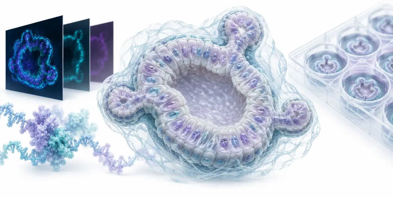
1. Morphology and Architecture: What Has Actually Formed?
Brightfield imaging provides an efficient way to monitor organoid formation and growth. Depending on the model, useful measurements include:
- Organoid number and formation efficiency.
- Size distribution, projected area, and growth over time.
- Circularity, budding, branching, and structural complexity.
- Lumen formation, wall thickness, and structural integrity.
These measurements should reflect the biology of the model. Budding can be informative in intestinal organoids, whereas other systems may require assessment of tubular organization, regional patterning, or chamber formation.
2. Cellular Identity and Composition: Are the Right Cells Present?
Cell identity should be established using a panel of complementary markers, interpreted alongside cellular location and morphology.
A single positive marker rarely establishes tissue identity, differentiation state, or functional maturity.
Illustrative marker panels
| Organoid model | Examples of markers to consider | What the panel helps assess |
|---|---|---|
| Intestinal | CDX2, VIL1, MUC2, CHGA; LGR5 when relevant | Intestinal identity and selected absorptive, secretory, and stem-cell populations |
| Hepatic or hepatobiliary | HNF4A, ALB; KRT19 for biliary components | Hepatocyte-associated identity and the presence of biliary populations |
| Neural | SOX2 or PAX6 for relevant progenitors; MAP2 and RBFOX3/NeuN for neuronal populations | Progenitor and neuronal composition in the intended regional context |
| Kidney | NPHS1 and WT1; LRP2; SLC12A1 | Selected podocyte, proximal tubule, and thick ascending limb-associated populations |
3. Tissue-Specific Function: Does the Model Perform the Required Task?
Functional assays connect an organoid’s identity to its intended application.
The most informative endpoint measures the process central to the research question. Detecting a transporter, enzyme, hormone, or neuronal marker does not establish that the associated biological system functions adequately.
Match the assay to the tissue and application
| Model or application | Examples of functional readouts | Key interpretation point |
|---|---|---|
| Intestinal epithelial organoids | Forskolin-induced swelling in validated CFTR assay systems; permeability in suitable formats | Swelling and barrier measurements assess different processes |
| Hepatocyte-containing organoids | Albumin secretion, urea production, CYP activity and inducibility | Select endpoints appropriate to the hepatocyte component and study objective |
| Neural organoids | Calcium imaging, patch-clamp recording, multielectrode array measurements | Calcium dynamics and electrical recordings provide complementary information |
| Kidney organoids | Uptake assays in identified tubular populations; selected transport assays | Uptake does not demonstrate whole-kidney filtration |
| Islet-like organoids | Glucose-stimulated insulin secretion and secretion dynamics | Insulin expression alone does not establish glucose responsiveness |
| Cardiac organoids | Contraction analysis, calcium handling, electrophysiological measurements | Spontaneous beating alone does not establish adult-like cardiac performance |
These approaches have experimental precedents. Forskolin-induced swelling was established as a functional CFTR readout in intestinal organoids, while hepatobiliary organoid studies have evaluated secretion and drug-metabolizing activity. Kidney organoid studies have demonstrated uptake within proximal tubule-like structures. Glucose-responsive insulin secretion has been demonstrated in human islet-like organoids, and cardiac organoid studies have combined contraction analysis with calcium imaging to assess functional behavior.
4. Growth, Viability, and Cell Death: Why Has the Signal Changed?
Organoid size, metabolic activity, proliferation, and survival are related but distinct endpoints.
A treatment can slow proliferation without killing cells. It can also alter cellular metabolism before membrane integrity changes.
Use complementary readouts
| Biological question | Example readouts | Main limitation |
|---|---|---|
| Are cells proliferating? | EdU incorporation, Ki-67 staining, longitudinal cell-number measurements | Labeling efficiency and the denominator require validation |
| How much metabolically active tissue remains? | ATP-based viability assays | ATP content can change independently of cell number |
| Is membrane integrity compromised? | Membrane-impermeant dyes, LDH release | Penetration, background, and sampling can affect results |
| Is apoptosis occurring? | Caspase activity, cleaved caspase-3, appropriately controlled Annexin V assays | No single endpoint captures every stage or mode of death |
ATP-based assays are widely used in three-dimensional cultures. Their signal reflects ATP associated with metabolically active cells, and assay formulations designed for 3D samples improve lysis of complex structures. Nevertheless, lysis efficiency and signal performance should be verified for the actual organoid size and matrix conditions.
A lower ATP signal alone does not prove cytotoxicity. Pair it with imaging, proliferation measurements, or a cell-death endpoint when that distinction affects the conclusion. Spatial analysis can also be valuable. A whole-well measurement may conceal a viable outer layer surrounding a damaged center.
5. Disease Phenotypes and Drug Responses: Is the Model Capturing the Biology of Interest?
Once baseline identity and function are established, phenotyping should focus on the intended disease mechanism or treatment effect.
Disease modeling
Useful endpoints may include:
- Altered lineage composition or tissue organization.
- Defective secretion, transport, or electrophysiological behavior.
- Abnormal proliferation or differentiation.
- Changes in inflammatory signaling or extracellular matrix deposition.
- Mechanism-specific molecular changes.
Strong experimental designs connect the observed phenotype to a plausible mechanism. Depending on the model, this may involve genetic correction, an isogenic comparator, a targeted perturbation, or pharmacological rescue.
Drug screening
For treatment studies, consider a panel that combines:
- A primary response endpoint, such as viable tissue burden or restoration of a defined function.
- An independent confirmation endpoint, such as structural imaging or cell-death analysis.
- A mechanistic endpoint, when needed to explain the response.
Report concentration, exposure duration, baseline conditions, normalization method, and dose–response fitting approach. An IC₅₀ is conditional on these choices; it is not an intrinsic property of a compound across all organoid assays.
When baseline growth rates differ substantially, endpoint comparisons can be confounded. Growth-rate-based approaches may help when their assumptions and measurement requirements are satisfied.
For patient-derived tumor organoids, confirm tumor identity and relevant molecular features. Avoid assuming that conventional epithelial cultures retain the original immune and stromal compartments.
6. How to Build a Practical Organoid Phenotyping Panel
The goal is to obtain enough complementary evidence to support the intended conclusion.
A practical starting panel
For many studies, an initial panel can include:
- Quantitative morphology at defined time points.
- A tissue-appropriate cell-identity panel.
- Baseline viability and proliferation measurements.
- At least one functional endpoint relevant to the intended claim.
- Appropriate controls and independent biological replication.
This is a planning framework, not a universal qualification standard. Establishing a new model may require much deeper characterization than applying an existing, well-characterized model to a narrow question.
7. Experimental Details That Make Phenotyping More Reliable
Control starting conditions
Document organoid size distribution, seeding density, passage number, culture age, medium composition, and extracellular matrix source or lot. These variables can influence baseline growth and treatment responses.
Validate measurements in three dimensions
Check reagent penetration, lysis efficiency, imaging depth, and assay interference. Include matrix-only or reagent-background controls where relevant.
For image analysis, keep acquisition settings consistent, validate segmentation, and examine representative failures. A brightfield projected area should not be reported as measured three-dimensional volume.
Distinguish biological from technical replication
Several wells from one culture preparation assess technical variability. They do not replace independent donors, cell lines, or differentiation batches.
Define the experimental unit before analysis. Multiple organoids within one well—and multiple wells from one donor—may require a statistical approach that accounts for their shared origin.
Normalize to the question
Secreted protein per well measures total output. The same protein normalized to viable cell number estimates output relative to viable tissue burden. Both can be informative, but they answer different questions.
Choose the denominator deliberately and check that treatment does not invalidate it.
Build a More Informative Organoid Study With Creative Bioarray
Selecting a phenotyping panel is easier when model development and downstream analysis are planned together.
Creative Bioarray offers Organoid Development Services covering organoid generation, culture, expansion, and characterization. Its Analytical & Readout Services include three-dimensional imaging, morphometric analysis, functional assays, molecular profiling, and bioinformatics support.
Whether you are establishing a model, investigating a disease phenotype, or evaluating treatment responses, discuss your tissue system and research objective with Creative Bioarray to develop an analysis strategy that connects organoid structure, cellular identity, and biological function.
Your email address will not be published. Required fields are marked *